Bone Densitometry

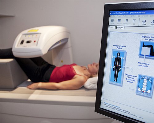
Bone density scanning, also called dual-energy x-ray absorptiometry (DXA) or bone densitometry, is an enhanced form of x-ray technology that is used to measure bone loss. DXA is today's established standard for measuring bone mineral density (BMD).
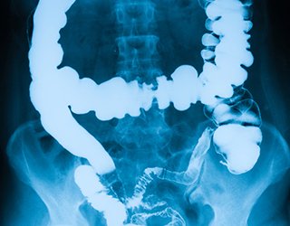
An x-ray (radiograph) is a noninvasive medical test that helps physicians diagnose and treat medical conditions. Imaging with x-rays involves exposing a part of the body to a small dose of ionizing radiation to produce pictures of the inside of the body. X-rays are the oldest and most frequently used form of medical imaging.
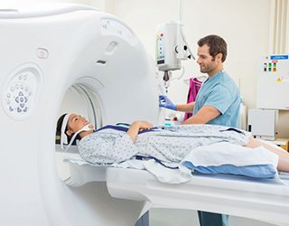
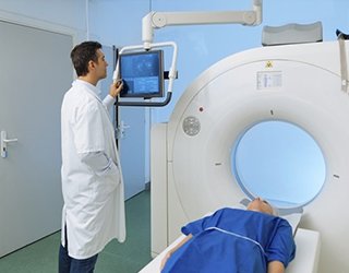
DXA is most often performed on the lower spine and hips. In children and some adults, the whole body is sometimes scanned. Peripheral devices that use x-ray or ultrasound are sometimes used to screen for low bone mass. In some communities, a CT scan with special software can also be used to diagnose or monitor low bone mass (QCT). This is accurate but less commonly used than DXA scanning.
An x-ray (radiograph) is a noninvasive medical test that helps physicians diagnose and treat medical conditions. Imaging with x-rays involves exposing a part of the body to a small dose of ionizing radiation to produce pictures of the inside of the body. X-rays are the oldest and most frequently used form of medical imaging.
DXA is most often performed on the lower spine and hips. In children and some adults, the whole body is sometimes scanned. Peripheral devices that use x-ray or ultrasound are sometimes used to screen for low bone mass. In some communities, a CT scan with special software can also be used to diagnose or monitor low bone mass (QCT). This is accurate but less commonly used than DXA scanning.
On the day of the exam you may eat normally. You should not take calcium supplements for at least 24 hours before your exam.
You should wear loose, comfortable clothing, avoiding garments that have zippers, belts or buttons made of metal. Objects such as keys or wallets that would be in the area being scanned should be removed.
You may be asked to remove some or all of your clothes and to wear a gown during the exam. You may also be asked to remove jewelry, removable dental appliances, eye glasses and any metal objects or clothing that might interfere with the x-ray images.
Inform your physician if you recently had a barium examination or have been injected with a contrast material for a computed tomography (CT) scan or radioisotope scan. You may have to wait 10 to 14 days before undergoing a DXA test.
Women should always inform their physician and x-ray technologist if there is any possibility that they are pregnant. Many imaging tests are not performed during pregnancy so as not to expose the fetus to radiation. If an x-ray is necessary, precautions will be taken to minimize radiation exposure to the baby. See the Safety page for more information about pregnancy and x-rays.
Bone density tests are a quick and painless procedure.
Routine evaluations every two years may be needed to see a significant change in bone mineral density, decrease or increase. Few patients, such as patients on high dose steroid medication, may need follow-up at six months.
Routine evaluations every two years may be needed to see a significant change in bone mineral density, decrease or increase. Few patients, such as patients on high dose steroid medication, may need follow-up at six months.
A radiologist, a physician specifically trained to supervise and interpret radiology examinations, will analyze the images and send a signed report to your primary care or referring physician, who will discuss the results with you.
DXA scans are also interpreted by other physicians such as rheumatologists and endocrinologists. A clinician should review your DXA scan while assessing the presence of clinical risk factors such as:
Your test results will be in the form of two scores:
- Rheumatoid Arthritis
- Chronic Renal and Liver disease
- Respiratory disease
- Inflammatory Bowel disease
Your test results will be in the form of two scores:
- T score — This number shows the amount of bone you have compared with a young adult of the same gender with peak bone mass. A score above -1 is considered normal. A score between -1 and -2.5 is classified as osteopenia (low bone mass). A score below -2.5 is defined as osteoporosis. The T score is used to estimate your risk of developing a fracture.
- Z score — This number reflects the amount of bone you have compared with other people in your age group and of the same size and gender. If this score is unusually high or low, it may indicate a need for further medical tests.
Other Guidlines

Children's (Pediatric) Voiding Cystourethrogram
Computed Tomography
(CT) - Body
Magnetic Resonance Imaging (MRI) - Body

Magnetic Resonance, Functional (fMRI) - Brain
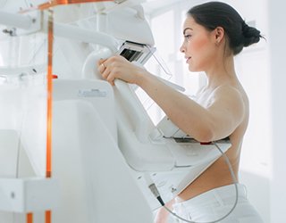
Mammography
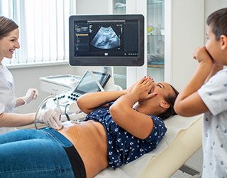
Obstetric Ultrasound
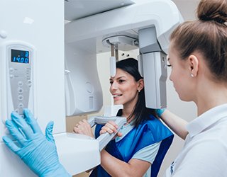
Panoramic Dental X-ray
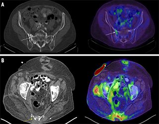
Positron Emission Tomography - Computed Tomography (PETCT)
Radionuclide
Ultrasound - Abdomen

Ultrasound-Guided Breast Biopsy