Children's (Pediatric) Voiding Cystourethrogram


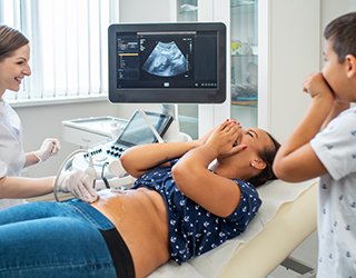
A children's (pediatric) voiding cystourethrogram (VCUG) is an x-ray examination of a child's bladder and lower urinary tract that uses a special form of x-ray called fluoroscopy and a contrast material.
An x-ray (radiograph) is a noninvasive medical test that helps physicians diagnose and treat medical conditions. Imaging with x-rays involves exposing a part of the body to a small dose of ionizing radiation to produce pictures of the inside of the body. X-rays are the oldest and most frequently used form of medical imaging.
Fluoroscopy makes it possible to see internal organs in motion. When the bladder is filled with and then emptied of a water-soluble contrast material, the radiologist is able to view and assess the anatomy and function of the bladder and lower urinary tract.
A voiding cystourethrogram enables a radiologist, a physician specifically trained to supervise and interpret radiology examinations, to detect abnormalities in the flow of urine through the lower urinary tract. This examination is often recommended after a urinary tract infection to check for a condition known as vesicoureteral (VU) reflux.
About VU Reflux
Urine is produced in the kidney and flows through the ureter, the tube that carries urine from each kidney to the bladder. A valve mechanism prevents urine from backing up into the kidneys as the bladder gets full. Urine leaves the bladder through the urethra and is eliminated from the body during urination.
In some children, an abnormality in the valve or the ureters allows urine to flow backwards, a condition called VU reflux. In mild cases urine backs up into the lower ureter. In severe cases it can back up into the kidney. Usually, children with this condition are born with it. Other causes include:
- Bladder obstruction
- Abnormal urination with very high pressure within the bladder
- Incomplete emptying of the bladder
- Urinary tract infection.
Urinary tract infection may be the only symptom of the problem.
You should inform your physician of any medications your child is taking and if he or she has any allergies, especially to contrast materials. Also inform your doctor about recent illnesses or other medical conditions.
Your child does not need to fast or wear special clothing. Explain to your child what will happen during the examination so that there will be no confusion about what is expected. Your child will have to remove all clothing and wear a gown.
Sedation is rarely needed.
Your child does not need to fast or wear special clothing. Explain to your child what will happen during the examination so that there will be no confusion about what is expected. Your child will have to remove all clothing and wear a gown.
Sedation is rarely needed.
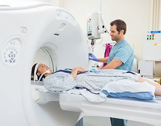
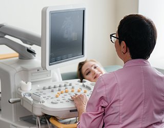
The equipment typically used for this examination consists of a radiographic table, one or two x-ray tubes and a television-like monitor that is located in the examining room. Fluoroscopy, which converts x-rays into video images, is used to watch and guide progress of the procedure. The video is produced by the x-ray machine and a detector that is suspended over a table on which the patient lies.
A catheter, a flexible, hollow plastic tube, will be used to fill the bladder with a water-soluble contrast material. The catheter has a diameter smaller than the urethra.
A catheter, a flexible, hollow plastic tube, will be used to fill the bladder with a water-soluble contrast material. The catheter has a diameter smaller than the urethra.
X-rays are a form of radiation like light or radio waves. X-rays pass through most objects, including the body. Once it is carefully aimed at the part of the body being examined, an x-ray machine produces a small burst of radiation that passes through the body, recording an image on photographic film or a special detector.
Fluoroscopy uses a continuous or pulsed x-ray beam to create a sequence of images that are projected onto a fluorescent screen, or television-like monitor. When used with a contrast material, which clearly defines the area being examined by making it appear dark (or by electronically reversing the image contrast to white), this special x-ray technique makes it possible for the physician to view joints or internal organs in motion. Still images or movies are also captured and stored either on film or electronically on a computer.
Until recently, x-ray images were maintained as hard film copy (much like a photographic negative). Today, most images are digital files that are stored electronically. These stored images are easily accessible and are frequently compared to current x-ray images for diagnosis and disease management.
This examination is usually done on an outpatient basis.
The technologist begins by positioning the child on the table. Infants and young children may be wrapped tightly in a blanket or other restraint to help them lie still during the imaging.
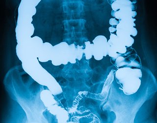
An x-ray of the abdomen may be performed before the urinary bladder is catheterized. The bladder catheterization is performed by a specially trained professional, a nurse, doctor or technologist. After cleaning the genital area, a catheter is inserted through the urethra, the tube that carries urine from the bladder out of the body. The catheter may be taped to the skin so that it will not be dislodged during the procedure. Then, the bladder is filled with a liquid contrast material. When the bladder is full, the child will urinate on the x-ray table. A urinal, bed pan or absorbent pad may be used to catch the liquid contrast material. The radiologist or technologist will use fluoroscopy to monitor the filling of the bladder and urination. X-ray images will be obtained during the monitoring. After the x-ray images are checked to make sure the exam is complete, the catheter is removed.
The radiologist will check to see if any of the liquid contrast material goes backward into one or both ureters and kidneys and whether the shape and contour of the bladder and urethra are normal.
You must hold very still and may be asked to keep from breathing for a few seconds while the x-ray picture is taken to reduce the possibility of a blurred image. The technologist will walk behind a wall or into the next room to activate the x-ray machine.
When the examination is complete, you will be asked to wait until the radiologist determines that all the necessary images have been obtained. A voiding cystourethrogram is usually completed within 30 minutes.
The technologist begins by positioning the child on the table. Infants and young children may be wrapped tightly in a blanket or other restraint to help them lie still during the imaging.
An x-ray of the abdomen may be performed before the urinary bladder is catheterized. The bladder catheterization is performed by a specially trained professional, a nurse, doctor or technologist. After cleaning the genital area, a catheter is inserted through the urethra, the tube that carries urine from the bladder out of the body. The catheter may be taped to the skin so that it will not be dislodged during the procedure. Then, the bladder is filled with a liquid contrast material. When the bladder is full, the child will urinate on the x-ray table. A urinal, bed pan or absorbent pad may be used to catch the liquid contrast material. The radiologist or technologist will use fluoroscopy to monitor the filling of the bladder and urination. X-ray images will be obtained during the monitoring. After the x-ray images are checked to make sure the exam is complete, the catheter is removed.
The radiologist will check to see if any of the liquid contrast material goes backward into one or both ureters and kidneys and whether the shape and contour of the bladder and urethra are normal.
You must hold very still and may be asked to keep from breathing for a few seconds while the x-ray picture is taken to reduce the possibility of a blurred image. The technologist will walk behind a wall or into the next room to activate the x-ray machine.
When the examination is complete, you will be asked to wait until the radiologist determines that all the necessary images have been obtained. A voiding cystourethrogram is usually completed within 30 minutes.
A voiding cystourethrogram may frighten some children. The antiseptic used to clean and prepare for the insertion of the catheter may feel cold. Some children may experience discomfort when the catheter is inserted and the bladder is filled with the liquid contrast material. Most children accept the procedure after an explanation of all of its parts.
A parent may be allowed to stay in the fluoroscopy room to comfort the child. Everyone, except the patient, wears a lead apron in the fluoroscopy room to protect from radiation exposure. A parent who wishes to remain in the fluoroscopy room will be required to wear a lead apron.
A parent may be allowed to stay in the fluoroscopy room to comfort the child. Everyone, except the patient, wears a lead apron in the fluoroscopy room to protect from radiation exposure. A parent who wishes to remain in the fluoroscopy room will be required to wear a lead apron.
A radiologist, a physician specifically trained to supervise and interpret radiology examinations, will analyze the images and send a signed report to your primary care or referring physician, who will discuss the results with you.
Follow-up examinations may be necessary, and your doctor will explain the exact reason why another exam is requested. Sometimes a follow-up exam is done because a suspicious or questionable finding needs clarification with additional views or a special imaging technique. A follow-up examination may also be necessary so that any change in a known abnormality can be monitored over time. Follow-up examinations are sometimes the best way to see if treatment is working or if an abnormality is stable over time.
Follow-up examinations may be necessary, and your doctor will explain the exact reason why another exam is requested. Sometimes a follow-up exam is done because a suspicious or questionable finding needs clarification with additional views or a special imaging technique. A follow-up examination may also be necessary so that any change in a known abnormality can be monitored over time. Follow-up examinations are sometimes the best way to see if treatment is working or if an abnormality is stable over time.
Benefits
- Voiding cystourethrograms provide valuable, detailed information to assist physicians in diagnosing and treating urinary tract conditions to prevent kidney damage.
- The examination results allow physicians to determine whether therapy is necessary. Some conditions require no therapy, while others may require medications. Some may even need surgery.
- No radiation remains in a patient's body after an x-ray examination.
- X-rays usually have no side effects in the typical diagnostic range for this exam.
- There is always a slight chance of cancer from excessive exposure to radiation. However, the benefit of an accurate diagnosis far outweighs the risk.
- The effective radiation dose for this procedure varies. See the Safety page for more information about radiation dose.
- Some children experience discomfort during urination immediately after the procedure. This discomfort usually resolves in less than 12 hours.
Risks
A Word About Minimizing Radiation Exposure
Special care is taken during x-ray examinations to use the lowest radiation dose possible while producing the best images for evaluation. National and international radiology protection organizations continually review and update the technique standards used by radiology professionals.
Modern x-ray systems have very controlled x-ray beams and dose control methods to minimize stray (scatter) radiation. This ensures that those parts of a patient's body not being imaged receive minimal radiation exposure.
A voiding cystourethrogram cannot evaluate obstruction of flow of urine from the kidneys. Additional examinations are needed if obstruction is suspected.
A voiding cystourethrogram should not be performed while an active, untreated urinary tract infection is present.
A voiding cystourethrogram should not be performed while an active, untreated urinary tract infection is present.
Other Guidlines

Bone Densitometry
Computed Tomography
(CT) - Body
Magnetic Resonance Imaging (MRI) - Body

Magnetic Resonance, Functional (fMRI) - Brain
Mammography
Obstetric Ultrasound
Panoramic Dental X-ray
Positron Emission Tomography - Computed Tomography (PETCT)
Radionuclide
Ultrasound - Abdomen
Ultrasound-Guided Breast Biopsy